

Dental disease (Periodontal Disease) is the most common disease that is diagnosed at Coxwell Animal Clinic. It is very common in both cats and dogs and can be preventable!
As young adults, most cats and dogs will begin to show signs of periodontal disease. This disease can go unnoticed by owners so it is important to check your pet’s mouth for signs of dental disease. Common signs are:
- Bad breath
- Loose or missing teeth
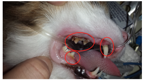
- Teeth covered in tartar or discolored
- Dropping food from the mouth
- Drooling
- Facial swelling or pain when touching mouth area
- Loss of appetite
Periodontal disease starts with bacteria build up on the surface of teeth, plaque. This plaque can harden into tartar and spread under the gum line. Once under the gums, the bacteria can spread toxins causing damage to the gums, teeth, and supporting tissues. This results in gingivitis, bones loss, and soft tissue loss. If left untreated your pet can suffer with the pain of dental disease and is at risk of losing teeth and having bacteria spread through their body
Prevention is the key factor in controlling periodontal disease! At a young age it is ideal to get your pet started on an oral care regime. This consists of:
- Brushing teeth
- Routine Check ups
- Appropriate Nutrition
- Please see our previous Blog Post on Dental Health https://www.coxwellvet.com/pet-dental-health/
If periodontal disease has begun and we are unable to control it with at home care, the next step would be to bring your pet in for a comprehensive dental procedure.
Before the procedure, we would be happy to see your pet for a pre-surgical exam and to perform a blood work to assess their suitability for anesthetic.
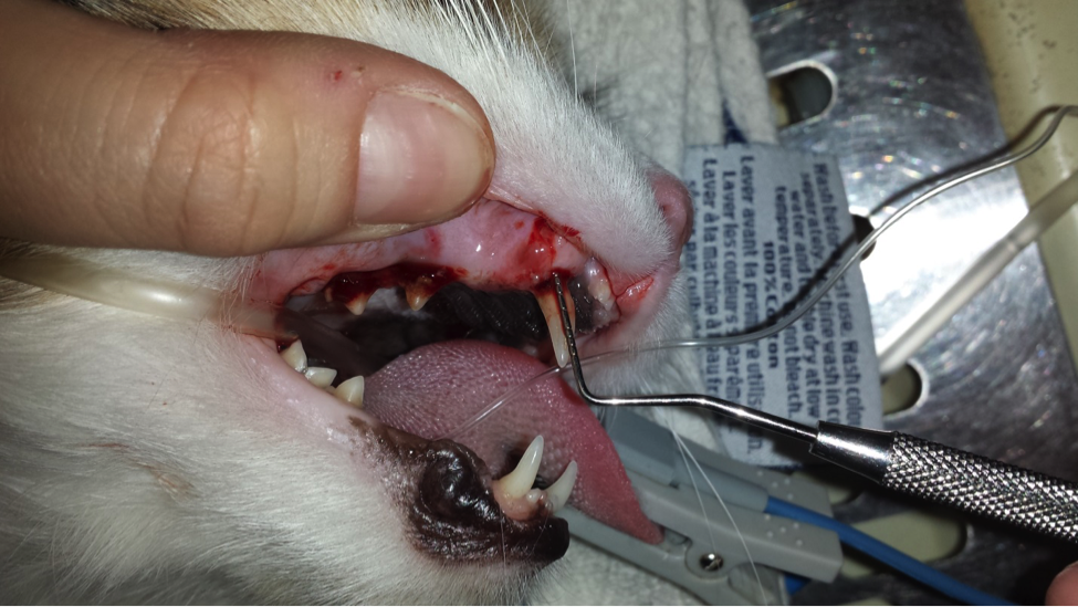
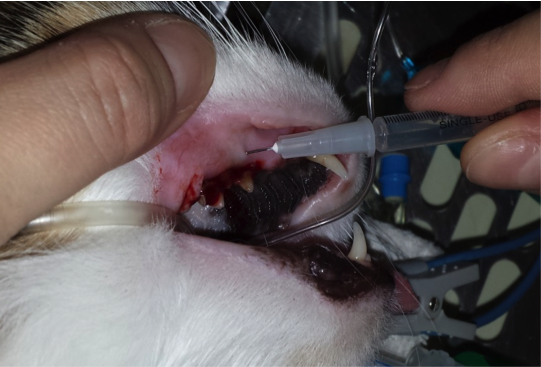
Once we have examined your pet, they are sedated and placed under general anesthetic. They are carefully monitored by our trained staff. Once stable the procedure begins; the following is a basic template of our dental procedures:
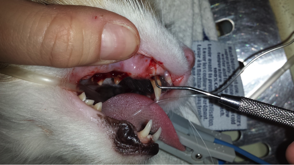
1. We perform a thorough exam of all teeth, check for bleeding, pockets around the roots, and chart all our findings.
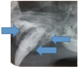
2. Once the oral cavity has been examined we perform full mouth x-rays. Quite often we may find a tooth that looks ok from the outside but the root may be diseased.

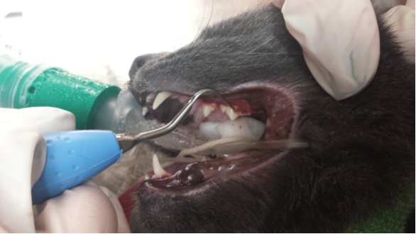
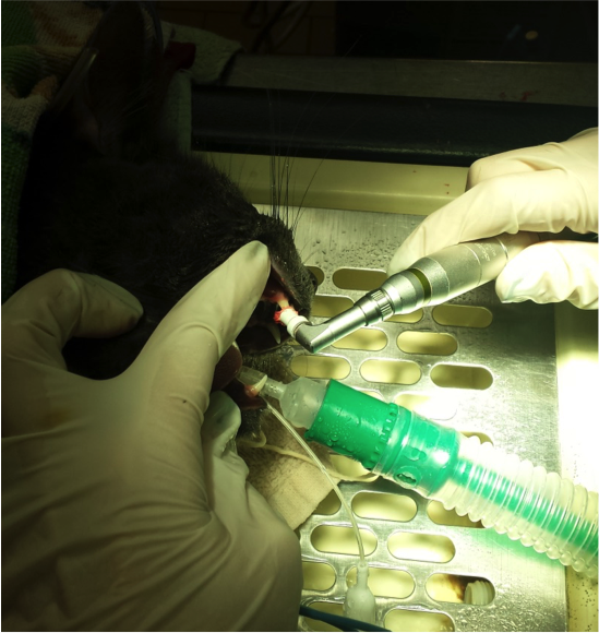
3. If there are any teeth that require extracting, they will be cleaned first and extracted. Prior to extractions there is local freezing administered to aid in pain control.
4. All remaining teeth are scaled and polished.
Once complete your pet will recover in hospital and sent home with dental care instructions. It is important to continue at home care following a dental to maintain a healthy mouth.
For more information please feel free to call us and book free a dental consultation appointment! Special thanks to Hazel and Gypsy for allowing us to take photos during their dental procedure.
Written by: Dr. Ashley Woo